DONATE
MEMBERSHIP
Menu
Animals
Plants
Wildlife Care
Saving Species
Wild and Fun
News
wildlife disease laboratories
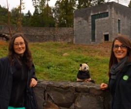
A Successful Giant Panda Workshop
July 4, 2015
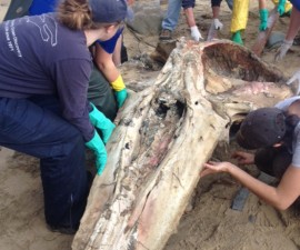
Bones on the Beach
May 7, 2015
Recommended
The Hatch of 2020
Wildlife Care